

More than three million years before ancient civilizations emerged, a small-brained, upright-walking hominin lived in what is now South Africa. Known as “Little Foot,” the fossil represents one of the most complete early human skeletons ever discovered. Scientists have now reconstructed the face of this ancient ancestor, using a particle accelerator.
Researchers say the remains belong to an early human relative in the genus Australopithecus, most likely the species Australopithecus prometheus. These ancient hominins lived long before modern humans appeared. They walked upright on two legs but still retained physical traits suited for climbing trees.
The skeleton was discovered in the Sterkfontein Caves in South Africa, a famous fossil site often called the “Cradle of Humankind.” The region has produced many discoveries that help scientists understand the early stages of human evolution.
Little Foot stands out because of its exceptional preservation. Many early hominin fossils consist of only a few bones or skull fragments. In contrast, this specimen includes a nearly complete skeleton, with bones from the skull, arms, legs, hands, and feet, allowing researchers to study early human anatomy in remarkable detail.
Fossil skull distorted by millions of years underground
The fossil was first identified in the 1990s when researchers discovered several small foot bones inside cave deposits. These bones later gave the skeleton its nickname. Excavating the rest of the fossil proved difficult because the fragile remains were embedded in hard rock.
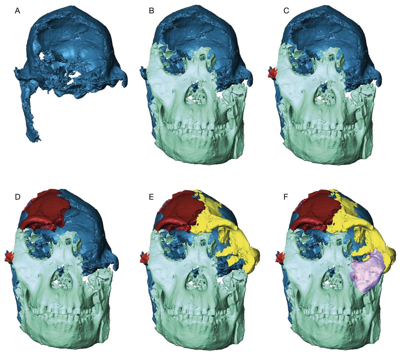
Over millions of years underground, geological pressure distorted the skull and shifted many bones from their original positions. When scientists completed the excavation, the facial bones were fragmented and displaced, making traditional reconstruction extremely difficult. Researchers needed a new approach to understand what the skull originally looked like.
Particle accelerator reveals hidden details
Scientists turned to a powerful synchrotron imaging facility known as the Diamond Light Source. This particle accelerator generates extremely intense X-rays capable of penetrating rock and fossil material.
Scientists have reconstructed the 3.7-million-year-old face of “Little Foot,” one of the most complete early human fossils ever discovered.
Using a particle accelerator, researchers digitally rebuilt the skull and revealed clues about how early Australopithecus ancestors looked. pic.twitter.com/Z8wskzFNli
— Tom Marvolo Riddle (@tom_riddle2025) March 6, 2026
Using this technology, researchers scanned the skull in extraordinary detail. The process produced more than 9,000 cross-sectional images. Each image captured a thin slice of the fossil, revealing structures hidden deep within the rock.
The scans allowed scientists to identify bone fragments, cracks, and distortions caused by geological pressure over millions of years.
Digital reconstruction rebuilds the ancient face
Researchers used specialized software to separate bone fragments from the surrounding rock layer by layer. They digitally repositioned sections of the skull that had shifted over time.
In areas where bones were too damaged, scientists mirrored intact parts from the opposite side of the skull to complete the reconstruction. This process produced a detailed digital model of Little Foot’s face.
Researchers then compared the reconstructed skull with those of modern humans, great apes, and other Australopithecus fossils. Scientists mapped anatomical reference points and measured facial dimensions to study similarities and differences.
Comparisons reveal surprising evolutionary links
The analysis revealed an unexpected pattern. Little Foot’s facial structure showed stronger similarities to an ancient fossil discovered in Ethiopia than to another skull found in the same South African cave system.
In several digital comparisons, the reconstructed face grouped more closely with the Ethiopian specimen known as A.L. 444-2 than with the southern African fossil Sts 5.
One notable feature is the shape of the eye sockets. Little Foot’s eye orbits are large and oval, resembling those seen in orangutans and the Ethiopian fossil. By contrast, the Sts 5 skull has more rectangular eye sockets.
Researchers say the orbit region varies across Australopithecus species and can provide clues about evolutionary changes in facial anatomy.
New clues about early human evolution
The findings suggest that some facial traits may have been shared among early Australopithecus populations living across eastern and southern Africa during the Pliocene epoch.
This suggests that ancient hominin groups across the continent were more interconnected than previously believed, perhaps through migration or shared ancestry.
Researchers caution that the reconstruction remains preliminary because fossils can become distorted after millions of years underground. Future studies using advanced modeling techniques may further refine the skull’s original shape.
Even so, the digital reconstruction marks an important step in understanding one of the earliest members of the human family. By combining particle accelerator imaging with modern digital reconstruction, scientists are uncovering new details about how early human ancestors looked and evolved across ancient Africa.

